


Lung Cancer Detection Using Artificial Intelligence
Posted by | Fuld & Company
Abstract
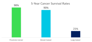
Lung cancer is the number one cause of cancer-related deaths, both in the United States and worldwide, killing three times as many men as prostate cancer and three times as many women as breast cancer. As reported by the American Cancer Society, the five-year survival rate after being diagnosed with lung cancer is just 21%, compared to 98% for prostate cancer and 90% for breast cancer.
Traditional Method of Cancer Diagnosis
- Image Detection: Chest CT scans contain images ranging from 200-to 500 slices, which are individually examined by a radiologist to detect cancerous nodules. These slices are difficult to detect with the naked eye and can result in false diagnoses.
- Diagnosis: Results show that only 68% of lung cancer nodules are correctly diagnosed when only one radiologist examines the scan, with up to 82% accurately detected by two radiologists. Each radiologist takes up to 3-4 minutes to look at scans and detect cancerous lung nodules.
- Accuracy: Accuracy has always been a concern with traditional diagnosis methods. Many patients are diagnosed with cancer, but after going through treatment, it turns out that they were misdiagnosed. These errors lead to misallocation of resources, and in some cases, even death.
Artificial Intelligence Approach to Cancer Diagnosis
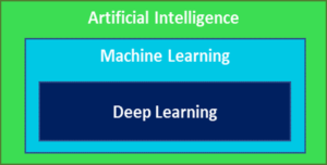
- Artificial Intelligence: AI enables the machine to make its own decisions without human intervention. In short, AI makes a computer behave the way humans do.
- Machine Learning: ML a subset of Artificial Intelligence, uses statistical algorithms to build systems that can automatically learn, improve from experiences without being explicitly programmed, and predict what is going to happen in the future. is.
- Deep Learning: A subset of Machine learning, this helps a computer model filter the input data through layers to predict and classify information. In short, the algorithm is given raw data and decides for itself what features are relevant.
Lung Cancer Detection System
Fuld & Company collaborated with a new startup to build a detection system to discover lung cancer at an earlier stage. Below are the top features of the system:
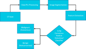
- Scanning: Reads all the MRI scans of the patient and creates a 3D structure from them.
- Data Pre-Processing:
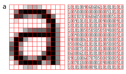
- Normalizes Pixel (pixel values are color code) values to 1 mm. [When images are passed through the neural network, the computation becomes complex. To reduce this complexity, pixels are normalized to 1.]
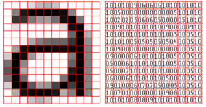
Figure: Converting an image into pixels (Source)
-
- Converts the pixels into Hounsfield Units. [The Hounsfield unit (HU) is a quantitative measurement of radio density used by radiologists in the interpretation of computed tomography (CT) images.] For Lungs, the HU threshold value is −700 to −600.
- Image Segmentation & Feature Extraction:
- Segments the region of interest (lungs) from the scans based on the HU of the system
- Extracts the features to find the cancerous nodule once the lung portion is segmented from the MRI scan
- Divides features into attributes that can help find the cancerous nodule. It can be texture features, or statistical features such as area, perimeter, centroid, diameter, or mean intensity.
- Classification
- Passes scans through the input layer of CNN and then passes through further various hidden layers, going through multiple iterations. When they reach the hidden layers, SoftMax functions are activated, and loss is computed using the SoftMax activation function. Cross-entropy is used as the loss function to be optimized. After multiple iterations and multiple optimizations, the system can identify the cancerous nodule.
Fuld & Co. along with the start-up team trained the system using a database of more than 50,000 CT scans which consisted of both cancer patients and non-cancer patients.
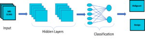
Figure: CNN Architecture
Accuracy
- Sensitivity: 0.90 (90%) – % of patients with cancer who are diagnosed as having cancer by the system.
- Specificity: 0.96 (96%) – % of patients who have not had cancer and are diagnosed as not having cancer by the system.
- Accuracy: 0.93 (93%) – Overall accuracy of the system.
Advantages of using our AI-enabled system
- Increased Radiologists’ ability to accurately detect cancerous nodules from 68% to more than 93%.
- Decreased time-to-detect from almost 2 minutes to seconds.
- Incorporated easily into the existing computer-aided diagnosis system.
- Decreased chances of false-positive cases to 1-2%.
Author – Sanidhya Singh


